Upper Inner Thigh Anatomy : Hip Pain Overcoming And Preventing It. The fascial compartments of thigh are the three fascial compartments that divide and contain the thigh muscles.the fascia lata is the strong and deep fascia of the thigh that surrounds the thigh muscles and forms the outer limits of the compartments. A person may feel pain in the inner thigh muscles or adductors. People who play soccer have these specific muscles of the leg very well defined, so they're like a walking anatomy atlas for thigh muscles. Human muscles · july 20, 2016. The adductor brevis, adductor longus and adductor magnus make up the the starting position is lying on the right side where the upper body is supported by the right arm.
In this condition, damage to a nerve that travels down the thigh (lateral femoral cutaneous nerve) causes burning, tingling, and numbness in the front and outer thigh. It's the area that runs from the hip to the knee in each leg. Medial muscles adduct and rotate your thigh, and posterior flex your leg and extend your thigh. All the medial thigh muscles are innervated by the obturator nerve, which arises from the lumbar plexus. Learn vocabulary, terms and more with flashcards, games and other study tools.
Groin Pain Structures And Conditions That Can Contribute To Groin Pain from mk0hippainhelp9h8quy.kinstacdn.com Internally the muscle compartments are divided by the lateral and medial intermuscular septa. They have a lot to do with how your hips move. The thigh muscles don't just move your legs. Lump on the inner thigh is a condition in which the skin on inner thigh roughens and forms either large or small bumps. Each compartment has a distinct innervation and function. This is because the anatomy of the thigh is complex and difficult to manipulate. Muscle anatomy of upper thigh. The thigh is the region between the hip and knee joints.
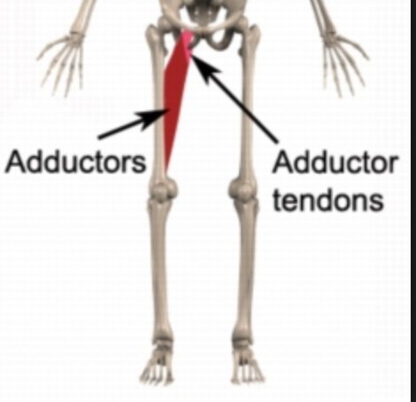
The adductor group, commonly known as the inner (upper) thighs, is a group of several muscles that, when engaged, move the legs together.
The adductor brevis, adductor longus and adductor magnus make up the the starting position is lying on the right side where the upper body is supported by the right arm. Like the forearm, the upper leg, or thigh, has a dense arrangement of many muscles. Posterior compartment, also known as the flexor compartment; This bone is very thick and strong and makes a ball and socket joint at the hip, and a hinge joint at the knee. The following diagram illustrates the actions of the terms adduction, abduction, flexion and extension at the different joints. These bumps can be of any size small or. The anatomy of the leg consists of those parts of the lower limb between the knee and the ankle. Human muscles · july 20, 2016. Sartorius muscle anatomy page has origin, insertion, innervation, and blood supply information. It features two bones known as the tibia, or shin bone, and the smaller fibula.depending on their location in the anatomy of the leg, its muscle groups are divided into four different regions called compartments. Each compartment has a distinct innervation and function. Learn vocabulary, terms and more with flashcards, games and other study tools. It is part of the lower limb.
The thigh is the region between the hip and knee joints. The rectus femoris is located in the center of the thigh, while the vastus medialis is in the middle of the said body part. Muscle anatomy of upper thigh. Started in 1995, this collection now contains 6952 interlinked topic pages divided into a tree of 31 specialty books and 737 chapters. Each compartment has a distinct innervation and function.
Hip And Thigh Bones Joints Muscles Kenhub from thumbor.kenhub.com Then roll to the outside of your foot. The longest tendon in the human body is the plantaris tendon, measuring between 30cm and 45cm. Learn vocabulary, terms and more with flashcards, games and other study tools. Upper inner thigh anatomy : Groin is the area of the actual junction of the inner thigh and torso including the genitals.crotch is more. Schreiber a person with defined leg muscles. The thigh bears much of the load of the body's weight when a person is upright. The musculature of the thigh can be split into three sections;
Thigh liposuction is interesting because it is one of the most common areas of liposuction performed, yet i think it is the worst area executed by surgeons.
The femoral nerve supplies the skin around the inner leg and the upper thigh area, the saphenous nerve supplies the medial aspect of the lower leg—a small area on of the foot and the ankle. Lump on the inner thigh is a condition in which the skin on inner thigh roughens and forms either large or small bumps. All the medial thigh muscles are innervated by the obturator nerve, which arises from the lumbar plexus. It also weakens the muscles in the upper thigh and can cause pain in the thigh near the knee or hip. You should feel the inner upper thigh tone or activate slightly. The tensor fasciae latae, or tfl, is a small muscle that runs from the side of your hip and down to the outer top part of your thigh. The anatomy of the leg consists of those parts of the lower limb between the knee and the ankle. The thigh is the area between the hip and the knee joint. The nerve damage may occur due to tight clothing, prior surgery, or pregnancy, certain repetitive excercise, or it may occur without a clear cause. Meanwhile, the vastus lateralis is on the side of the thigh, while the vastus intermedius is hidden below the rectus femoris(5). The longest tendon in the human body is the plantaris tendon, measuring between 30cm and 45cm. Groin is the area of the actual junction of the inner thigh and torso including the genitals.crotch is more. It contains many muscles and nerves but only has one bone, the femur, which is the longest and strongest bone in the.
Groin is the area of the actual junction of the inner thigh and torso including the genitals.crotch is more. Repetitive stress on your thigh muscles can lead to inflammation, pain, and swelling of the tendon, known as tendonitis. The thigh bears much of the load of the body's weight when a person is upright. The thigh (proximal lower limb) muscles are arranged into three compartments : The musculature of the thigh can be split into three sections;
How To Treat Adductor Tendonitis The Art Of Manliness from content.artofmanliness.com The following diagram illustrates the actions of the terms adduction, abduction, flexion and extension at the different joints. When doing inner thigh lifts keep your upper body stable, your core tight and maintain the working leg straight and the foot flexed. In this condition, damage to a nerve that travels down the thigh (lateral femoral cutaneous nerve) causes burning, tingling, and numbness in the front and outer thigh. The hip muscles are going to be slip into hip muscles and gluteal muscles. Ebraheim's educational animated video describes muscle anatomy of the thigh. This is because the anatomy of the thigh is complex and difficult to manipulate. People who play soccer have these specific muscles of the leg very well defined, so they're like a walking anatomy atlas for thigh muscles. Pain in the groin and.
On the anterior side, the most prominent of the muscles are the sartorius muscle and the four muscles that make up quadriceps muscle group (the quads.)
Medial muscles adduct and rotate your thigh, and posterior flex your leg and extend your thigh. The hip muscles are going to be slip into hip muscles and gluteal muscles. People who play soccer have these specific muscles of the leg very well defined, so they're like a walking anatomy atlas for thigh muscles. The fascial compartments of thigh are the three fascial compartments that divide and contain the thigh muscles.the fascia lata is the strong and deep fascia of the thigh that surrounds the thigh muscles and forms the outer limits of the compartments. The adductor brevis, adductor longus and adductor magnus make up the the starting position is lying on the right side where the upper body is supported by the right arm. A person may feel pain in the inner thigh muscles or adductors. This automatically brings better balance to the inner and outer thighs. This bone is very thick and strong and makes a ball and socket joint at the hip, and a hinge joint at the knee. They have a lot to do with how your hips move. The thigh is the area between the hip and the knee joint. The single bone in the thigh region is called the femur. It transmits the great saphenous vein, and other, smaller vessels, and is termed the fossa ovalis. Medial compartment, also known as adductor compartment;
Then roll to the outside of your foot upper thigh anatomy. This automatically brings better balance to the inner and outer thighs.

0 Comments:
Posting Komentar