Home
Uncategories
Human Bone Anatomy Spine - The Vertebral Column Joints Vertebrae Vertebral Structure - On anatomical parts the user can choose between three types of labels:
Human Bone Anatomy Spine - The Vertebral Column Joints Vertebrae Vertebral Structure - On anatomical parts the user can choose between three types of labels:
Human Bone Anatomy Spine - The Vertebral Column Joints Vertebrae Vertebral Structure - On anatomical parts the user can choose between three types of labels:. There are anatomical variations both in skeletal and nonskeletal components in all these regions. 1 your spine in this region has a natural inward curve. On series the user can select the radiographs concerning the spine as a whole, the cervical, thoracic and lumbar vertebrae, the sacrum and coccyx. Spine or vertebral column | spine bones joints | human spine anatomy 3d animation | elearninthis video illustrates one of the main parts of human body, the s. See lumbar spine anatomy diagram stock video clips.
Shop for human spine anatomy at walmart.com. The disks that cushion vertebrae may compress with age or injury, leading to a herniated disk. Vertebrae separated by intervertebral discs. The vertebral column is a part of the axial skeleton, which comprises the skull, ribs and sternum other than the vertebral column. Standard radiographic view of anatomical structures of the spinal column.
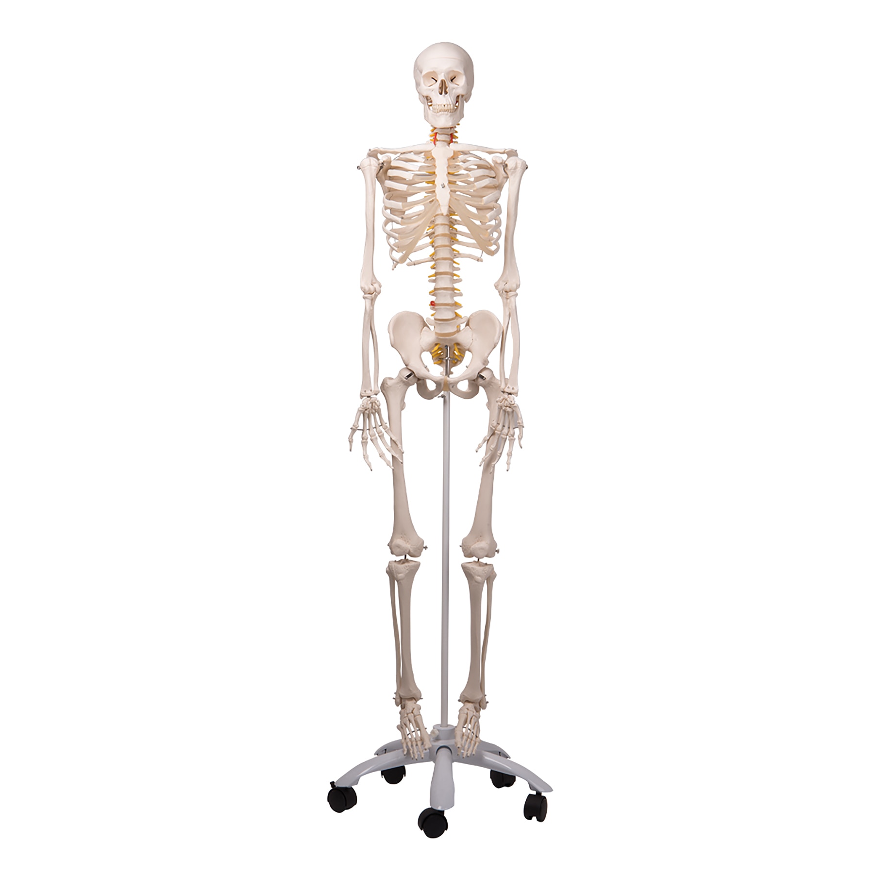
Amazon Com Evotech Disarticulated Human Skeleton Model For Anatomy 67 Inch High Full Size Skeleton Models With 3 Poster Skull Spine Bones Articulated Hand Foot For Anatomy Medical Learning Everything Else from images-na.ssl-images-amazon.com The spine supports your body and helps you walk, twist and move. Posterior view of the lumbar spine and pelvis. Buy anatomy spine at amazon. The meninges surround both brain and spinal cord and are filled with a liquid. The body creates two types of. It is composed of 300 bones at birth, but later decreases to 80 bones in the axial skeleton and 126 bones in the appendicular skeleton. There are anatomical variations both in skeletal and nonskeletal components in all these regions. These sections are cervical (neck), thoracic (upper and middle back), lumbar (lower back), and sacrum (tailbone).
This protects the spinal cord inside.
Protecting the spinal cord and nerves structural support for the body, allowing us to stand upright. 1 your spine in this region has a natural inward curve. Buy anatomy spine at amazon. See lumbar spine anatomy diagram stock video clips. Facet joints connect each vertebra, with fluid supporting. This protects the spinal cord inside. Spinal vertebrae bone spine vertebra toracica spinal cord spine structure back diagram spine sections spinal cord vertebrae spinal structure health diagram. Continue scrolling to read more below. 4 the si joints are located on either side of the sacral spine and are situated deep in the pelvis. The vertebral column is a part of the axial skeleton, which comprises the skull, ribs and sternum other than the vertebral column. The human skeleton, like that of other vertebrates, consists of two principal subdivisions, each with origins distinct from the others and each presenting certain individual features.these are (1) the axial, comprising the vertebral column—the spine—and much of the skull, and (2) the appendicular, to which the pelvic (hip) and pectoral (shoulder) girdles and the bones and cartilages of the. Together, the brain and spinal cord make up the central nervous system. The lumbar spine is composed of five vertebrae, named l1 to l5 from superior to inferior.
It is composed of 300 bones at birth, but later decreases to 80 bones in the axial skeleton and 126 bones in the appendicular skeleton. The spine or backbone consists of 26 small bones or vertebrae. On series the user can select the radiographs concerning the spine as a whole, the cervical, thoracic and lumbar vertebrae, the sacrum and coccyx. These sections are cervical (neck), thoracic (upper and middle back), lumbar (lower back), and sacrum (tailbone). Each si joint is secured and well protected by strong ligaments.
Human Skeleton With Flexible Spine Model Health Edco from www.healthedco.com The meninges surround both brain and spinal cord and are filled with a liquid. It is composed of 300 bones at birth, but later decreases to 80 bones in the axial skeleton and 126 bones in the appendicular skeleton. On series the user can select the radiographs concerning the spine as a whole, the cervical, thoracic and lumbar vertebrae, the sacrum and coccyx. Posterior view of the lumbar spine and pelvis. Each si joint is secured and well protected by strong ligaments. This spinal column provides the main support for your body, allowing you to stand upright, bend, and twist, while protecting the spinal cord from injury. Your lower back (lumbar spine) is the anatomic region between your lowest rib and the upper part of the buttock. Vertebrae separated by intervertebral discs.
Vertebrae separated by intervertebral discs.
The spine is composed of 33 bones called vertebrae, which stack together to form the spinal canal. This curve, called lordosis, helps to: Cores of marrow in the heads of long bones create about 500 billion red blood cells per day in hematopoiesis. Posterior view of the lumbar spine and pelvis. The meninges surround both brain and spinal cord and are filled with a liquid. The lumbar spine is composed of five vertebrae, named l1 to l5 from superior to inferior. On series the user can select the radiographs concerning the spine as a whole, the cervical, thoracic and lumbar vertebrae, the sacrum and coccyx. Together, the brain and spinal cord make up the central nervous system. The human spine is composed of 4 sections of vertebrae. Buy anatomy spine at amazon. The pelvis is composed of the two pelvic bones and the sacrum and coccyx (the pelvic bones are also known as the coxal, innominate, or hip bones) (fig. The human skeleton, like that of other vertebrates, consists of two principal subdivisions, each with origins distinct from the others and each presenting certain individual features.these are (1) the axial, comprising the vertebral column—the spine—and much of the skull, and (2) the appendicular, to which the pelvic (hip) and pectoral (shoulder) girdles and the bones and cartilages of the. The spine or backbone consists of 26 small bones or vertebrae.
These sections are cervical (neck), thoracic (upper and middle back), lumbar (lower back), and sacrum (tailbone). The meninges surround both brain and spinal cord and are filled with a liquid. The spine is made of 33 individual bones stacked one on top of the other. The human spine is a complex anatomic structure that is the scaffolding for the entire body. The lumbar and sacrum region make up the bone of the lower back anatomy.
3d Illustration Of Spinal Cord Thoracic Vertebrae A Part Of Human Skeleton Anatomy The Vertebral Column Also Known As The Canstock from cdn.w600.comps.canstockphoto.com It is composed of 300 bones at birth, but later decreases to 80 bones in the axial skeleton and 126 bones in the appendicular skeleton. The pelvis is composed of the two pelvic bones and the sacrum and coccyx (the pelvic bones are also known as the coxal, innominate, or hip bones) (fig. Free shipping on qualified orders. Two views of the vertebral column: Made up of 34 bones, the spinal column holds the body upright, allows it to bend and twist with ease and provides a conduit for major nerves running from the brain to the tips of the toes—and everywhere in between. Balance the weight of your head on top of your spine evenly distribute weights from your upper body into the lower extremities Understanding lower back anatomy is key to understanding the root of lower back and hip pain. Using this atlas of human anatomy of the spine and back.
Spine can be divided into cervical, thoracic, lumbar, sacral and coccygeal regions.
Strong muscles and bones, flexible tendons and ligaments, and sensitive nerves contribute to a healthy spine. Together, the brain and spinal cord make up the central nervous system. Made up of 34 bones, the spinal column holds the body upright, allows it to bend and twist with ease and provides a conduit for major nerves running from the brain to the tips of the toes—and everywhere in between. The disks that cushion vertebrae may compress with age or injury, leading to a herniated disk. The spine or backbone consists of 26 small bones or vertebrae. These sections are cervical (neck), thoracic (upper and middle back), lumbar (lower back), and sacrum (tailbone). On anatomical parts the user can choose between three types of labels: On series the user can select the radiographs concerning the spine as a whole, the cervical, thoracic and lumbar vertebrae, the sacrum and coccyx. Standard radiographic view of anatomical structures of the spinal column. Thoracic & lumbar vertebrae poster showing most common characteristics of the vertebrae along with ligament attachments to the ribs. Continue scrolling to read more below. Spine can be divided into cervical, thoracic, lumbar, sacral and coccygeal regions. This curve, called lordosis, helps to:
This protects the spinal cord inside human bone anatomy. Using this atlas of human anatomy of the spine and back.


0 Comments:
Posting Komentar